 HYANNIS – October is Breast Cancer Awareness Month, and for women living on the Cape there is good and bad news. First the bad news: Cape Cod continues to have the highest rates of breast cancer in the Commonwealth of Massachusetts. The good news is that Cape Cod Healthcare is bringing the latest diagnostic technology right to your doorstep.
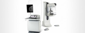
Tomosynthesis, or 3D mammography, is coming in early 2016. And for breast cancer detection, it’s a game changer.
3D mammography is similar to traditional two-dimensional (2D) mammography, using X-rays and compression to capture breast tissue images. Unlike 2D technology, the 3D machine actually moves around the compressed breast. Similar in concept to a CT scan, it takes many more pictures at different angles, providing a greater depth of view. The images are much clearer and show subtle differences that might have been previously hidden from detection.
With increased sensitivity, 3D mammography has greater accuracy in determining the size, shape and location of abnormalities. It also makes it easier to distinguish more serious cancers from less non-invasive ones like ductal carcinoma in situ (DCIS).
“Studies have demonstrated that 3D mammography increases the detection rate of cancer, while reducing the number of false positives,” said Salvatore Viscomi, MD, Chairman of the Department of Radiology. “It picks up the cancers we want to pick up, at a much earlier time, which is our goal. ”
Traditional screening mammography up to this point has had an undeniable role in reducing the death rate from breast cancer. However, it has limitations and produces too many false positive findings and anxiety provoking callbacks.
How much better is 3D technology? A study published in a 2014 edition of the Journal of the American Medical Association (JAMA) summarized the work of investigators seeking answers to that question. After reviewing data collected from 13 different sites across the country, they observed a 29 percent increase in the overall cancer detection rate despite a 15 percent decrease in recalls. In other words, fewer women returned for additional screening which revealed a greater number of cancers.
There are limitations in the design of this study, which was a retrospective comparison of the two different types of scans. However, investigators are confident in the findings, as they have been replicated in European studies as well.
Understanding the breast cancer statistics on the Cape, Dr. Viscomi has high hopes for the impact that this technology will have on women’s lives.
3D mammography will be used as the primary breast cancer screening tool, eventually replacing all of the 2D machines currently in use. It may also be used for callbacks and to assist in biopsies.
But, mammography is not the only technology currently in use and on the horizon. Ultrasound and MRI are also used routinely for cancer screening or diagnosis. Most often, they are used when warranted after follow-up to an initial mammogram. They are also used to guide biopsies.
The decision on which technology to use and when is very specific to the patient’s breast health history, genetic risk, or diagnosis, according to Dr. Viscomi. Different situations call for different approaches and there is no one size fits all model.
In the future, he envisions an expansion in the use of screening MRI. Currently, it is reserved for:
• women who have an elevated risk due to family history or genetics
• patients already diagnosed with cancer to look for additional cancers
• a better understanding of the nature of a detected abnormality
• women who have dense breast tissue.
MRI screening also has one important advantage: it eliminates the uncomfortable breast compression. Scans are done while lying prone on a table.
Dr. Viscomi predicts that with advancements in the strength and speed of MRI technology, it will eventually become the new standard screening tool. “The cost of the test is coming down, there is no exposure to radiation, and it is superior in detection,” he said.


Speak Your Mind